Figure 3.
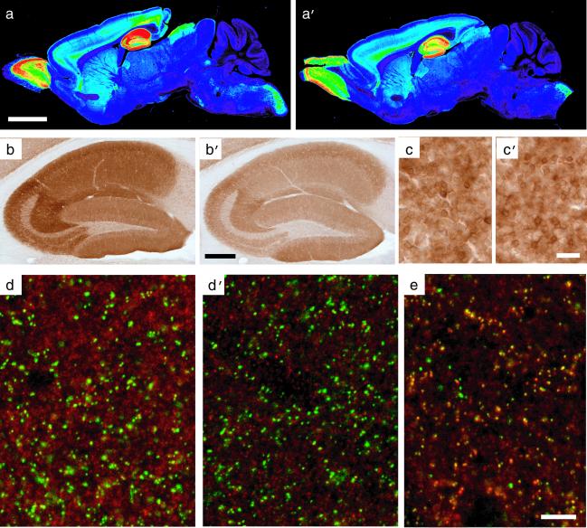
Regional and cellular expression of the α5-subunit protein. Selective loss of extrasynaptic α5GABAA receptors in α5(H105R) mice [a, b, c, and d: wild type; a′, b′, c′, d′, and e: α5(H105R) mutant]. (a and a′) False-color images depicting the regional distribution of the α5-subunit in parasagital sections of adult mice, as detected by immunoperoxidase staining. Staining in wild type corresponded to that described (11, 31). Note the reduction of staining selectively in the hippocampal formation of mutant mice. (b and b′) Enlargement of the hippocampal formation showing the global reduction of α5-subunit immunoreactivity in CA1 and CA3 in the mutant compared with control. (c and c′) By comparison, no change in α5-subunit staining intensity was observed in olfactory bulb granule cells (see Table 1 for quantification). (d and d′) Images from confocal laser scanning microscopy, depicting a double-immunofluorescence staining for the α5-subunit (red) and gephyrin (green) in the stratum radiatum of CA1. Gephyrin is a postsynaptic marker of GABAergic synapses. The lack of colocalization with the α5-subunit staining reflects the extrasynaptic distribution of α5GABAA receptors. The staining intensity of these receptors is markedly reduced in mutant mice, whereas gephyrin immunoreactivity is unchanged. (e) For comparison, double staining for the α2 subunit (red) and gephyrin (green) reveals the extensive postsynaptic localization of α2GABAA receptors (yellow dots) in α5 mutant mice. (Scale bars: a, a′, 2.5 mm; b, b′, 0.5 mm; c, c′, 25 μm; d, d′, and e, 10 μm.)

