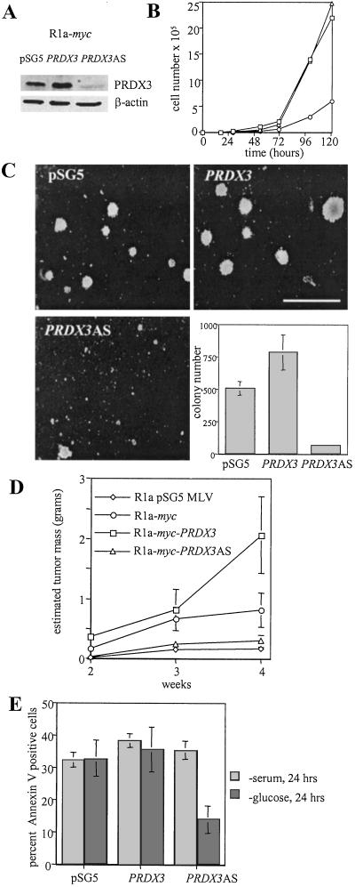
Figure 2.
Effect of PRDX3 expression on doubling time, transformation, and apoptosis in R1a-myc cells. (A) Immunoblot analysis of cell lysates from R1a-myc cells transfected with pSG5 empty vector, pSG5-PRDX3, or pSG5-PRDX3AS. (B) Growth curves of R1a-myc transfectants: pSG5 (□), PRDX3 (▵), and PRDX3AS (○). Doubling times were 10.4, 10.9, and 19.0 h, respectively. (C) Photomicrographs of methylcellulose colonies. (Bar = 500 μM.) The bar graph represents the average colony number per 35-mm dish ± SD. (E) Tumor formation in nude mice. The average estimated tumor mass was plotted at 2, 3, and 4 weeks after injection ± SD (n = 8). (D) Percentage of apoptotic cells 24 h after serum deprivation (light bars) or glucose deprivation (dark bars). The average ± SD of three experiments is shown.

