Figure 1.
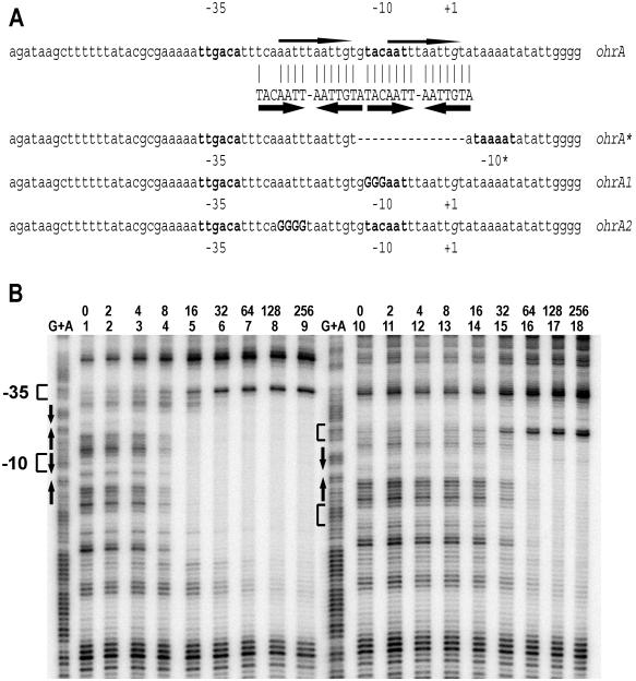
Binding of OhrR to the ohrA and ohrA* promoters. (A) The ohrA promoter and variants used in this study. The wild-type promoter (ohrA) contains two inverted repeats shown by thick arrows. Bases matching the perfect inverted repeat are identified by a vertical line. The thin arrows indicate the 11-bp direct repeats. The −10 and −35 regions are shown in bold letters, and the transcription start site (+1 position) is in italics. ohrA* is a previously described 15-bp deletion in the promoter region (10) as indicated with a dashed line. The ohrA1 mutation (uppercase bold letters) destroys the perfect inverted repeat (but not the direct repeat). The ohrA2 mutation (uppercase bold letters) destroys the direct repeat as well as the imperfect inverted repeat. −10* is a new −10 region in the ohrA* promoter. (B) DNase I footprinting analysis of OhrR binding to the ohrA (lanes 1–9) and ohrA* (lanes 10–18) promoters. Both promoter regions (2 nM DNA) were labeled on the top strand, and the amount of OhrR (nM) is shown above the lane number. Note that at high concentrations the protein binding site extends in the downstream direction. DTT (1 mM) was present in all binding reactions. The −10, −35, and +1 positions are shown next to the G+A ladders. The inverted repeats are shown with arrows.

