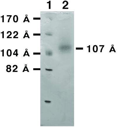
Figure 1.
Native polyacrylamide gradient gel electrophoresis of rHDL/CYP2B4. Dialysate containing rHDL and CYP2B4 was run on an 8–25% gradient gel and stained with Coomassie brilliant blue. The sizes of the rHDL structures were estimated from the known hydrodynamic diameters of the protein standards.

