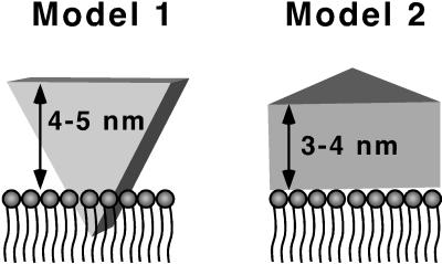
Figure 6.
Schematic of P450 interacting with the phospholipid bilayer domain of rHDL. In Model 1, a hydrophobic tip is inserted into the membrane and the heme is perpendicular to the membrane. In Model 2, the enzyme is lying on its distal face with the heme parallel to the membrane. The transmembrane anchor domain is not shown.

