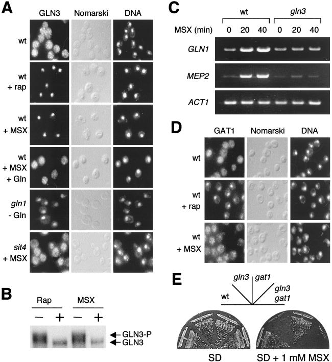
Figure 2.
Glutamine starvation specifically activates the transcription factor GLN3 but not GAT1. (A) Localization of GLN3-myc (GLN3) in a wild-type (wt) (TB123) and gln1 (JC33–6c) and sit4 (TB136–2a) mutant cells. Wild-type cells were grown in SD and treated either with rapamycin (+ rap) or MSX (+ MSX) for 20 min. Wild-type cells also were treated with MSX for 20 min and then with glutamine (5 mM final concentration) for 5 min (+ MSX + Gln). Untreated wild-type cells were used as a control. gln1 mutant cells were grown in SD supplemented with 0.3% glutamine and shifted to glutamine-free medium for 20 min. sit4 mutant cells were treated with MSX for 20 min. All strains were grown in SD at 30°C. Cells and DNA were visualized by Nomarski optics and 4′,6-diamidino-2-phenylindole staining (DNA). (B) GLN3 is dephosphorylated in glutamine-starved cells. Wild-type cells (TB123) were grown in SD and treated with either rapamycin or MSX for 25 min. GLN3-myc was detected by immunoblotting. (C) Reverse transcriptase–PCR analysis of total RNA from wild-type (JK9–3da) and gln3 (TB103–1d) mutant cells with oligonucleotide primers specific to the actin gene (ACT1) and two nitrogen-regulated genes, GLN1 and MEP2. Cells were grown in SD to an OD600 of 0.5 and treated with MSX for either 20 or 40 min. Nontreated cells (time 0) were used as a control. (D) GAT1-HA (GAT1) was visualized by indirect immunofluorescence in wild-type (TB106–2a) cells untreated (Control) or treated with rapamycin (+ rap) or MSX (+ MSX) for 20 min. (E) Growth of wild-type (JK9–3da), gln3 (TB103–1d), gat1 (TB102–1a), and gln3 gat1 (TB105–3b) mutant cells in SD or SD supplemented with a sublethal concentration (1 mM) of MSX. Plates were incubated at 30°C for 3 days.

