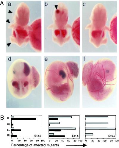
Figure 3.
Development of a severe bullous disorder in GRIP1−/− embryos. (A) Gross morphology of GRIP1−/− E12 embryos, showing large serous bullae on the surface of the head (a and b, arrowheads), upper and lower limbs (a) and with a large haemorrhagic bulla in the right lateral ventricle of the brain (b, arrowhead). In GRIP1−/− E14 embryos (shown inside the yolk sac), haemorrhagic bullae were found in similar locations: head (d and e), upper limbs (d). wt littermate controls are shown for comparison (c and f). (Magnifications: a–c, ×4; d–f, ×25.) (B) Frequency of serous (black bars) and haemorrhagic (gray bars) bullae in the head (H), upper limbs (UL), lower limbs (LL), and back bone (Bb) of GRIP1−/− embryos at different stages of embryonic development. The data derive from the cumulative observation of 35, 31, and four GRIP1−/− embryos at E12, E14, and E16, respectively.

