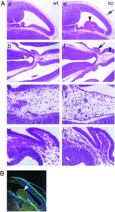
Figure 5.
Development of bullae in the lateral ventricle of the brain. (A) Hematoxylin-eosin staining of brain sections from wt (a–d) and GRIP1−/− (e–h) E12 embryos. Large haemorrhagic bulla (e, arrowhead) and blister on the surface of the meninges (e, arrow). A large serous blister (f, arrowhead; g, higher magnification) and extensive detachment of the neural epithelium from the underlying mesenchymal tissue (f, arrow; h, higher magnification) can be observed in the lateral ventricle, in close proximity to the developing choroid plexus. (B) PECAM immunostaining of a large bulla (arrowhead) protruding into the cerebral ventricle. Lack of PECAM staining on the surface of the bulla rules out its vascular origin. (Magnifications: A a, b, e, and f, ×20; c, d, g, and h, ×50; B, ×20.)

