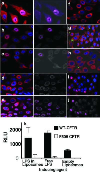
Figure 2.
In vitro translocation of NF-κB to the nucleus of cultured human epithelial cells expressing either ΔF508 or wt CFTR. Sections were stained for NF-κB (red) and nuclear DNA (blue). FITC-labeled LPS is green. Translocation of NF-κB into the nucleus is shown as a magenta color produced from overlap of the blue and red fluorescence signals in the images to the immediate right of a–e. (a) Wt CFTR cells incubated with P. aeruginosa whole bacteria. (b) Wt CFTR cells incubated with P. aeruginosa LPS in liposomes. (c) Wt CFTR cells incubated with free P. aeruginosa LPS. (d) Wt CFTR cells incubated with P. aeruginosa and CFTR peptide 108–117. (e) Wt CFTR cells incubated with P. aeruginosa and a scrambled version of CFTR peptide 108–117. (f) Wt CFTR cells incubated with E. coli LPS in liposomes. (g) ΔF508 CFTR cells incubated with wt P. aeruginosa FITC-LPS in liposomes. (h) Wt CFTR cells incubated with FITC-labeled P. aeruginosa incomplete core LPS (lacks CFTR ligand) in liposomes. (i) ΔF508 CFTR cells treated with 10% glycerol overnight to induce cell-surface expression of the ΔF508 CFTR protein, then incubated with wt P. aeruginosa LPS. (j) Colocalization depiction of i. (k) Induction of NF-κB luciferase activity, measured as relative light units (RLU), in transfected wt or ΔF508 CFTR cells exposed to P. aeruginosa LPS in liposomes or as free LPS. No activity above background was seen when ΔF508 CFTR cells were exposed to P. aeruginosa LPS.

