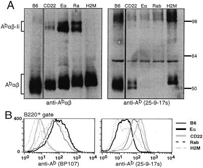
Figure 5.
Each peptide–Ab complex adopts a distinct conformation. (A) Abαβ dimers from splenocyte lysates of C57BL/6 (lane 1), CD22-dbl0 (lane 2), Eα-dbl0 (lane 3), Rab-dbl0 (lane 4), and H-2M0 (lane 5) mice were visualized by Western blotting of 8% SDS/PAGE gels. Blots were performed with rabbit antisera against the cytoplasmic tails of the Ab α and β chains (Left) or the conformation-dependent anti-Ab mAb 25-9-17s (Right). (B) Splenocytes from the same mice were stained with BP107 (Left) or 25-9-17s (Right) and analyzed by flow cytometry. The staining shown is on B220+-gated cells.

