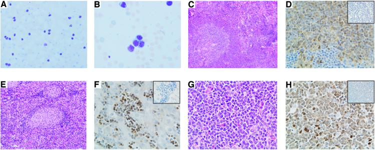
Figure 4.
Histopathological analysis of the Eμ-TCL1 mice. (A) Blood smear stained with Wright Giemsa showing an increased number of circulating lymphocytes. (B) High magnification of the blood smear. (C) Histology of spleen, liver (E), and cervical lymph node (G) after hematoxylin-eosin staining. (D) Immunodetection of Tcl1 protein in spleen, liver (F), and cervical lymph node (H). (Insets) Negative control in which the primary antibody has been omitted.

