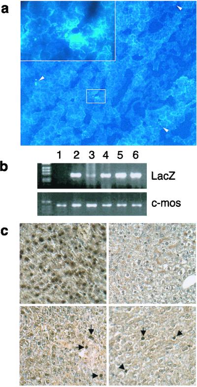
Figure 2.
Detection of transplanted hepatocytes in recipient liver. (a) Hoechst 33258-positive cells were detected in frozen liver sections from SCID recipient mice 8 days after intrasplenic transplantation. (Magnification, ×100.) In the upper left corner is a ×400 magnification of the selected area. (b) PCR analysis for LacZ gene or murine-specific c-mos protooncogene. Genomic DNA was extracted from the liver of SCID mouse (lane 1) and from the liver of donor HNF1-LacZ transgenic mice (lane 2). The LacZ sequence was detected in genomic DNA from transplanted SCID liver with LacZ hepatocytes (lanes 3 and 4) and HBx-LacZ hepatocytes (lanes 5 and 6). (c) In vivo detection of LacZ expression by immunohistochemical staining. (Upper Left) HBx-LacZ donor mouse liver (positive control). (Upper Right) Untransplanted SCID mouse liver (negative control). (Lower Left) SCID mouse liver transplanted with HBx-LacZ hepatocytes. (Lower Right) SCID mouse liver transplanted with LacZ hepatocytes.

