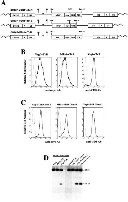
Figure 1.
Transduction of primary lymphocytes and cloned T cell lines by cTcR-expressing retroviral vectors. (A) Structure of retroviral vectors used in this study. VEGF-cTcR, VEGF chimeric TCR; MR-1-cTcR, MR-1 single-chain monoclonal antibody chimeric TCR; SD, splice donor; SA, splice acceptor; CMV IE, cytomegalovirus immediate early promoter; myc, human c-Myc epitope; CD8, CD8α hinge region; TCR, ζ chain of the TCR; LTR, long-terminal repeat. (B) FACS analysis of untransduced CTLs (dashed line) or CTLs transduced with VEGF-cTcR or MR-1-cTcR (solid line). Splenocytes transduced with VEGF-cTcR also were incubated with anti-CD8-FITC antibody (solid line) or an anti-rat IgG-FITC negative control antibody (dashed line). (C) FACS analysis of VEGF-cTcR clone 2 and MR-1-cTcR clone 8 cells. (D) Southern blot analysis of genomic DNA from transduced primary CD8+ splenocytes and CTL clones. Indicated is the expected 600-bp XbaI-BglII fragment containing the transgenic VEGF sequence and a background band that migrated as a 2.5-kb fragment. The one-copy control lane represents mock-transduced genomic DNA spiked with 12 pg of CMMP-VEGF-cTcR plasmid DNA, which correlates with an expected one copy of transgene per genome.

