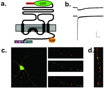
Figure 1.
Expression and functional analysis of FLAG-GFP-GluR2. (a) Schematic drawing of FLAG-GFP-GluR2 indicating the binding sites within the C-terminal domain for NSF and PDZ domain containing proteins. (b) Representative electrophysiological responses from HEK293 cells transfected with FLAG-GFP-GluR2(Q). Cells were held at −70 mV and kainate (1 mM, Upper) or glutamate (1 mM, Lower) was applied. Scale bars = 100 pA, 100 ms. (c) Surface labeling (red) indicates that a proportion of the total FLAG-GFP-GluR2 (green) is delivered to the cell surface. (d) FLAG-GFP-GluR2 (green) colocalizes with synaptophysin (red) in hippocampal neuronal cultures.

