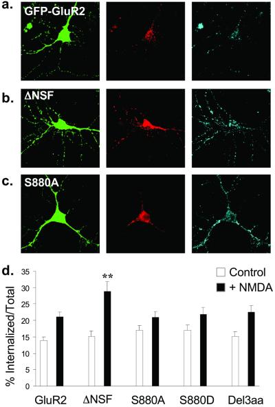
Figure 4.
NMDA induces endocytosis of FLAG-GFP-GluR2 that is regulated by interaction with NSF. (a–c) Representative images of transfected cells in which endocytosis of AMPARs was triggered by NMDA application. Panels are the same as in Fig. 3. (a) FLAG-GFP-GluR2. (b) FLAG-GFP-GluR2(ΔNSF). (c) FLAG-GFP-GluR2(S880A). (d) Quantification shows all constructs exhibit significant internalization (P < 0.05) after NMDA stimulation (filled columns) compared with control conditions (open columns). There is significantly increased NMDA-induced internalization of FLAG-GFP-GluR2(ΔNSF) (**, P < 0.01) compared with FLAG-GFP-GluR2. There is no significant difference between constructs in the absence of NMDA (open columns). ANOVA: df = 4, MS = 64.7, F = 1.21, P = 0.31.

