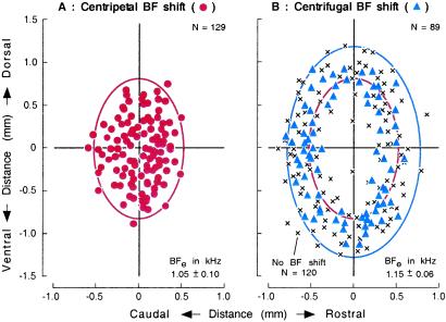
Figure 3.
Distributions of cortical neurons that exhibited centripetal or centrifugal BF shift. (A) Centripetal BF shift, ●. (B) Centrifugal BF shift, ▴ and no BF shift, ×. Locations of recorded neurons along the cortical surface are plotted relative to that of stimulated cortical neurons at the origin of the coordinates. x and y axes, directions parallel or orthogonal to the frequency axis of the AI. Data are pooled from 16 hemispheres of 11 animals. BFe, BF of electrically stimulated cortical neurons. Ellipse, area for centripetal or centrifugal BF shifts.

