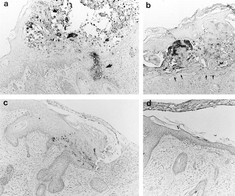
FIG. 1.

Histological analysis of VZV-infected skin implants. Subcutaneous skin implants infected with P-Oka (a), V-Oka (b), or gC−-Oka (c) or mock infected (d) were fixed in paraformaldehyde, paraffin embedded, and cut into 3-μm sections before in situ hybridization. Darkly stained cells indicate VZV DNA; tissue was counterstained lightly with hematoxylin. At day 21 postinfection, gC−-Oka lesions appeared in the epidermis (c). V-Oka lesions were larger; however, the basement membrane remained intact (arrows), and virus did not penetrate into the dermis (b). P-Oka skin lesions were largest (a), and VZV DNA was detected deep within dermal fibroblasts (arrow). Mock-infected skin had a normal appearance (d), and no DNA was detected in the implant. Histology shown is representative of four implants. Magnification, ×83.
