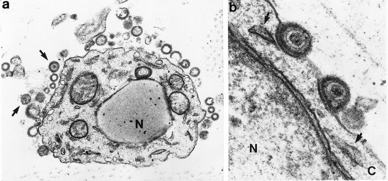
FIG. 2.

VZV release from skin cells, determined by transmission electron microscopy of skin cells 21 days after inoculation with VZV-S. (a) Enveloped virons containing dense cores have dispersed from the cell (arrows). Vacuoles from which particles egressed are visible adjacent to the cell membrane. Nucleocapsids are not visible in the nucleus (N) by this staining method. Magnification, ×24,000. (b) Higher-magnification view of virions with intact lipid bilayer on cell surface. Remnants of cytoplasmic vacuoles which carried virions to plasma membrane are indicated by arrows. N, nucleus; C, cytoplasm. Magnification, ×108,000.
