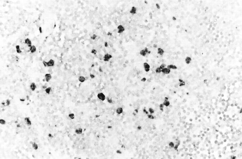
FIG. 7.
Histological analysis of an HSV-infected thy/liv implant inoculated with HSV-1 KOS and harvested on day 2 after infection. The implant was fixed in paraformaldehyde, paraffin embedded, and cut into 3-μm sections before in situ hybridization was performed. Darkly stained cells indicate where HSV-1 DNA was detected in cortical epithelial cells. Uninfected T cells were lightly counterstained with hematoxylin. Magnification, ×131.

