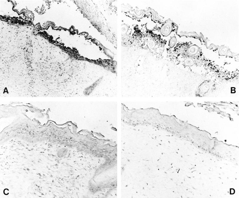
FIG. 8.

Histological analysis of HSV-infected skin implants. Subcutaneous skin implants were inoculated with HSV-1 KOS (A), HSV-1 ΔgC2-3rev (B), or HSV-1 ΔgC2-3 (C) or mock infected (D) and harvested on day 6 postinfection. Implants were fixed in paraformaldehyde, paraffin embedded, and cut into 3-μm sections before in situ hybridization was performed. HSV-1 DNA was detected in the equivalent epidermal lesions produced by KOS and ΔgC2-3rev (A and B). No HSV-1 DNA was detected in the epidermis of ΔgC2-3- or mock-infected implants (C and D). Hematoxylin counterstain revealed tissue histology. Magnifications: A and B, ×61; C and D, ×122.
