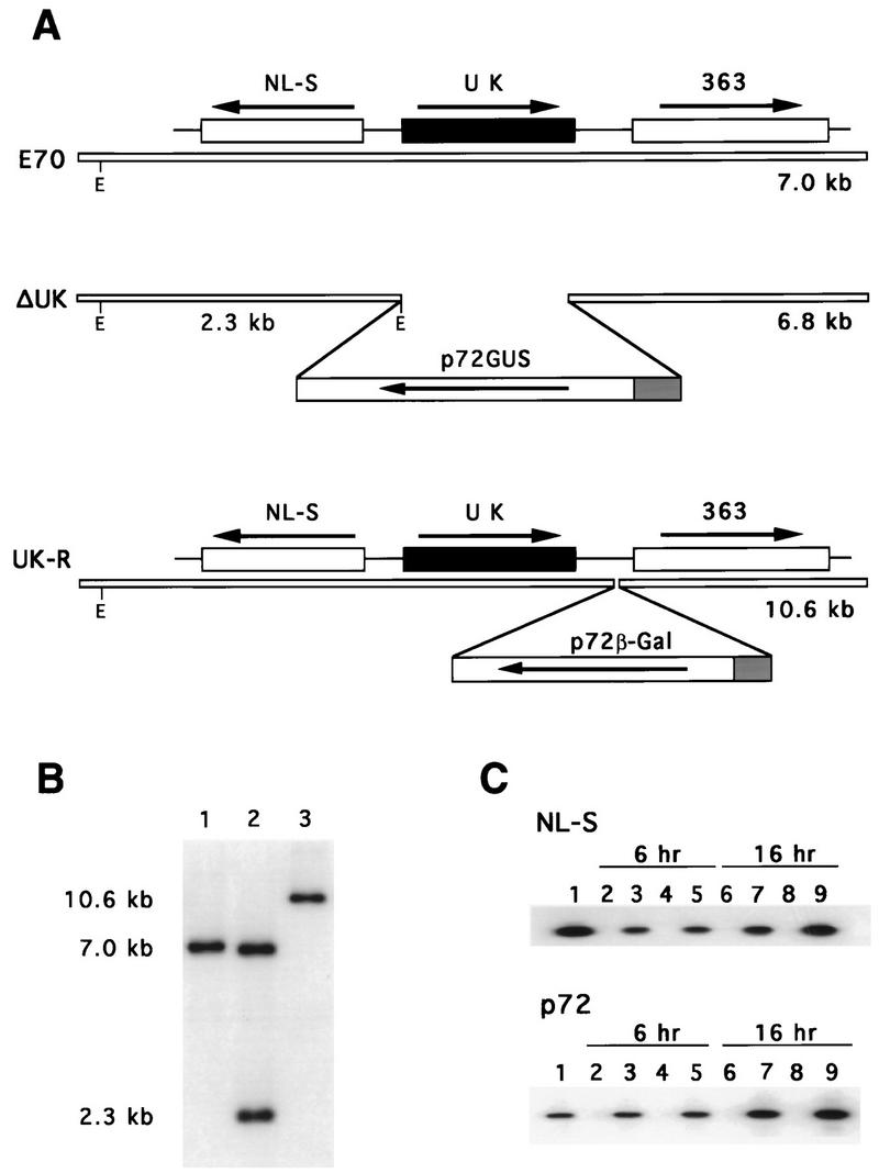
FIG. 3.

Characterizations of an ASFV UK gene deletion mutant, ΔUK, and its revertant, UK-R. (A) Diagram of the UK gene regions in the parental E70 isolate, the deletion mutant, ΔUK, and its revertant, UK-R. E, EcoRI site. (B) Southern blot analysis of E70 (lane 1), ΔUK (lane 2), and UK-R (lane 3). Purified viral DNAs were digested with EcoRI, electrophoresed, blotted, and hybridized with a DNA probe including UK gene sequences and flanking regions. Positions of molecular size markers are shown in kilobase pairs at the left. (C) RT-PCR amplification at 6 and 16 h postinfection of RNAs from macrophages infected with E70 (lanes 2, 3, 6, and 7) and ΔUK (lanes 4, 5, 8, and 9). One hundred nanograms of total RNA was used in the assay with either NL-S gene-specific or p72 gene-specific primers (lanes 3, 5, 7, and 9). PCR amplification from genomic DNAs (lane 1) and non-reverse-transcribed, DNase-treated RNA samples from macrophages infected with E70 (lanes 2 and 6) and ΔUK (lanes 4 and 8) were included as controls.
