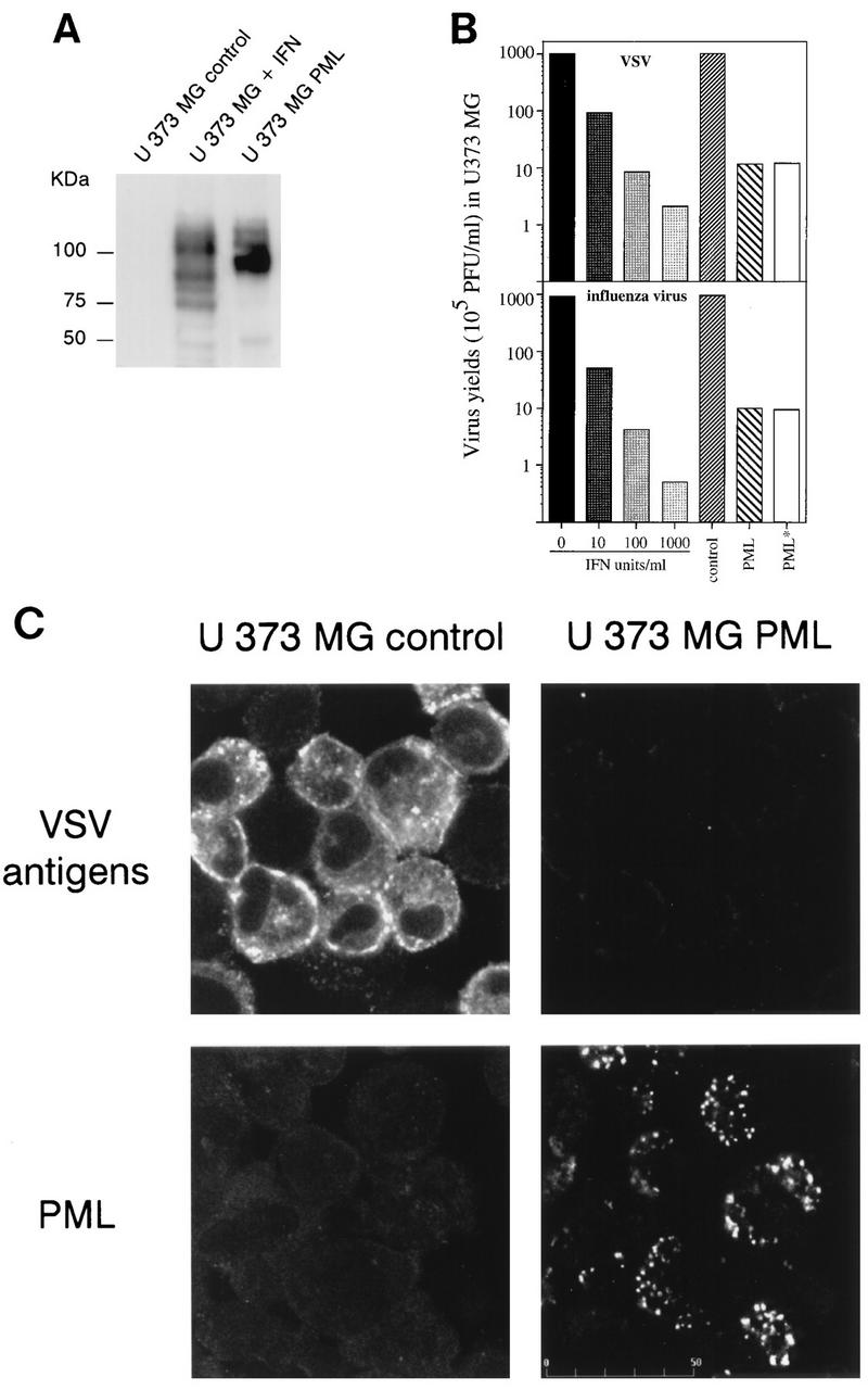
FIG. 3.

(A) PML level in IFN-treated U373 MG cells and U373 MG PML. U373 MG cells were treated for 48 h with 1,000 U of IFN-β per ml. Samples (50 μg) of extracts of control IFN-treated cells and U373 MG PML were analyzed by Western blotting and revealed with rabbit anti-PML antibodies. Note that all bands visualized by anti-PML antibodies are likely to be isoforms derived from alternative splicing of unique gene. Molecular size markers are indicated on the left. (B) Inhibition of virus replication in IFN-treated U373 MG cells and U373 MG PML. One series of U373 MG cells was treated for 48 h with 10, 100 or 1,000 U of IFN-β per ml. The second series of cells, U373 MG control (transfected with the empty vector), U373 MG PML, and U373 MG PML* (+ anti-IFN-α/β/γ antibodies [see Materials and Methods]) was seeded at 37°C for 5 h. The two series were then infected with VSV or influenza A virus at an MOI of 0.1. After 16 h, viral titers were determined as described in Materials and Methods. (C) Expression of PML in U373 MG cells inhibits the expression of VSV antigens (Top) Expression of VSV antigens in infected U373 MG control cells (transfected with the empty vector) and U373 MG PML. Immunofluorescence with rabbit anti-VSV antibodies was performed 13 h after infection with VSV at an MOI of 0.1 and revealed by FITC labelling. (Bottom) Expression of PML in U373 MG and U373 MG PML cells revealed by immunofluorescence with mouse anti-PML antibodies visualized with Texas red.
