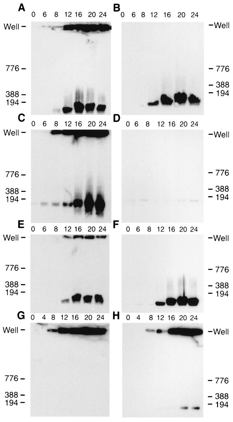
FIG. 6.

HSV-1 DNA analyzed by PFGE. Infected cells were harvested at the indicated times (hours) postinfection and treated as described in Materials and Methods. Following PFGE, the DNA was transferred to GeneScreen Plus and hybridized by using the HSV-1 BamHI K fragment (32P labeled) as a probe. Scanned images of the autoradiograph obtained from the Southern blots are shown. (A and B) Vero cells infected with KOS; (C and D) Vero cells infected with KUL25NS; (E and F) 8-1 cells infected with KUL25NS; (G) Vero cells infected with GCB; (H) C1 cells infected with GCB. (B, D, and F) Samples were treated with DNase prior to PFGE as described in Materials and Methods. Numbers next to each blot refer to molecular weight markers (kilobase pairs); the position of well DNA is marked for each blot.
