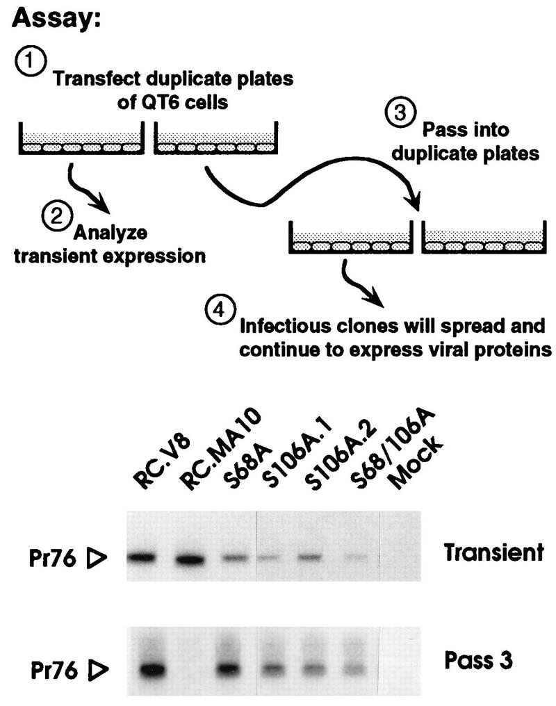
FIG. 3.

Qualitative analysis of infectivity. Each indicated clone was used to transfect duplicate plates of QT6 cells. One plate from each set was metabolically labeled for 2 h with [35S]methionine immediately following transfection, and the Gag proteins were collected by immunoprecipitation (Transient). The remaining plate of cells was passaged 2 days posttransfection and every 3rd day thereafter. On the 3rd day of the third passage, the cells were metabolically labeled with [35S]methionine, and again the Gag proteins were collected by immunoprecipitation (Pass 3). Proteins were separated by SDS-PAGE and visualized by fluorography. Only the region of the gel containing the Gag precursors is shown. RC.V8 and RC.MA10 are wild-type and negative controls, respectively. S106A.1 and S106A.2 are independent clones containing the same mutation.
