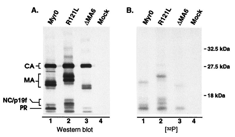
FIG. 5.
Analysis of pp12 species. 32P-labeled virions produced from infected TEFs were pelleted through a 25% sucrose cushion. Following suspension in sample buffer, the viral proteins were separated by SDS–12% PAGE and transferred to nitrocellulose. (A) Immunoblot with anti-RSV serum. (B) Autoradiograph of the same blot. R121L and ΔMA6 contain mutations which cause MA-related proteins to migrate either slower or faster, respectively, during electrophoresis. The locations of the wild-type and slower-migrating forms of p19f are indicated on the left side of panel A.

