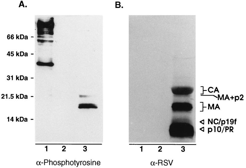
FIG. 6.
Detection of tyrosine-phosphorylated MA. (A) Proteins from sucrose gradient-purified RSV were separated by SDS–12% PAGE, transferred to nitrocellulose, and probed with an antiphosphotyrosine antibody and visualized by enhanced chemiluminescence. (B) Later, the same blot was stripped of antibodies and reprobed with anti-RSV serum. Lanes: 1, Src-transformed Rat-1 cell extract (positive control); 2, Rat-1 cell extract (negative control); 3, RSV.

