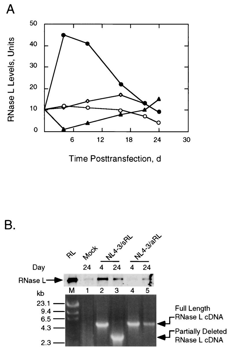
FIG. 3.

RNase L levels increase and then decline after transfection with NL4-3/sRL due to a deletion of RNase L coding sequence. (A) RNase L levels from Jurkat cells transfected with pNL4-3 (○), pNL4-3/Δnef (◊), pNL4-3/aRL (▴), or pNL4-3/sRL (•). The basal level of RNase L in untreated cells was an average of values obtained at 4, 16, and 24 days (d) of cell culture. (B) Upper panel, Western blot analysis of RNase L from transfected Jurkat cells probed with monoclonal antibody against human RNase L; lower panel, PCR amplification of the integrated RNase L cDNA. RL, 3 ng of RNase L; M, λ/HindIII molecular size markers (numbers to left are in kilobases). Plasmid names and times posttransfection in days are indicated.
