
FIG. 2.
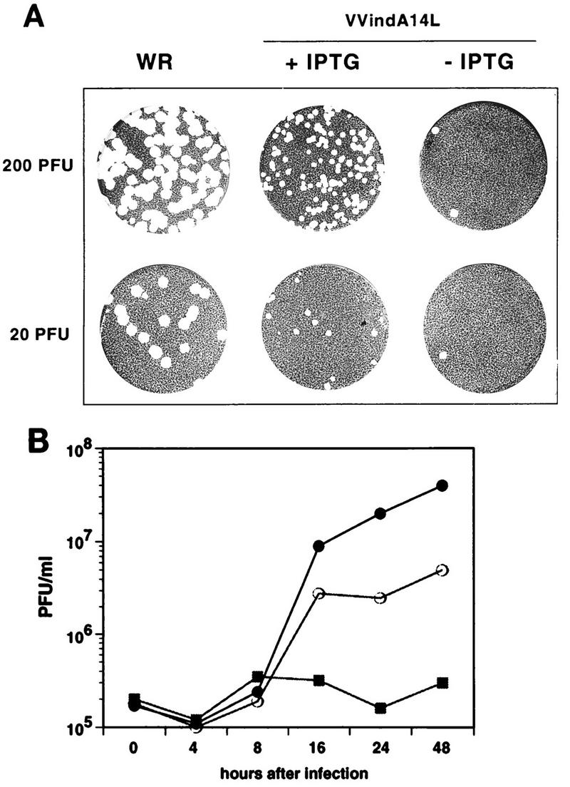
VVindA14L virus growth is dependent on the presence of IPTG. (A) Plaque assay. Confluent monolayers of BSC40 cells were infected with the indicated PFU of either WR or VVindA14L and overlaid with a mixture consisting of DMEM, 0.9% Bacto Agar, and 2% NCS, containing or lacking 2 mM IPTG. After 5 days, the monolayers were stained with 1% crystal violet. (B) One-step growth curves. BSC40 cells were infected at an MOI of 2.5 PFU/cell with WR virus (•) or with VVindA14L in the presence (○) or absence (▪) of 2 mM IPTG. Cells were collected at the indicated times after infection, and virus yields were determined by titration on BSC40 cells in the presence of 2 mM IPTG.