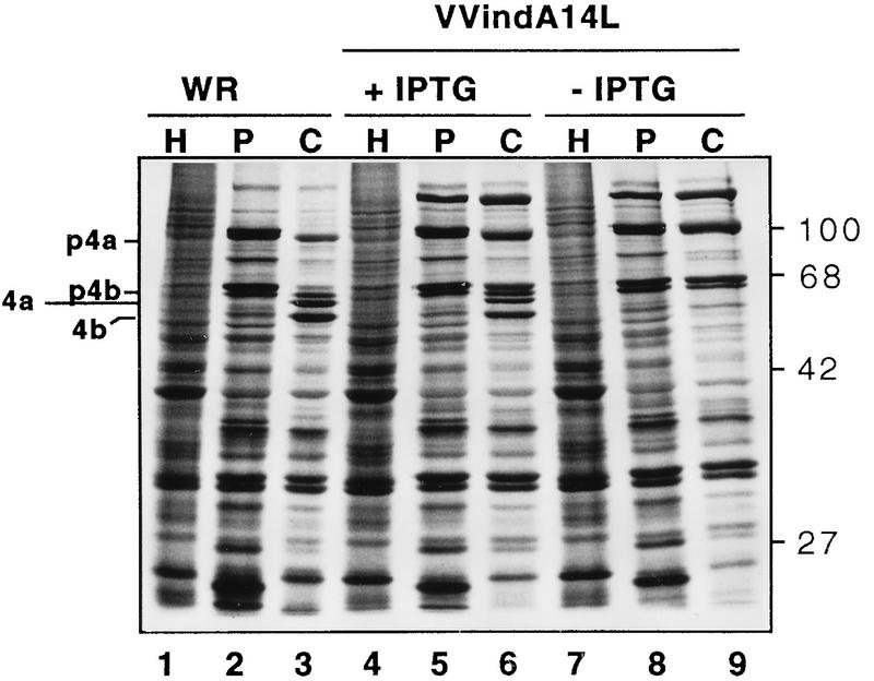
FIG. 4.
Synthesis and proteolytic processing of viral proteins in VVindA14L-infected cells. BSC40 cells were infected with WR (lanes 1 to 3) or with VVindA14L in the presence (lanes 4 to 6) or absence (lanes 7 to 9) of IPTG (2 mM). One culture of each group of infected cells was treated with HU (5 mM). At 6 hpi untreated and treated cells were pulse-labeled with [35S]methionine for 30 min and then chased with unlabeled methionine. The HU-treated cells (lanes H) and one culture of untreated cells from each group (lanes P) were harvested immediately after the start of the chase period, while the remaining untreated cells (lanes C) were kept in culture for another 18 h. Cells were lysed in sample buffer, and proteins were resolved by SDS-PAGE (12% polyacrylamide gel) and visualized after autoradiography of the dried gel. The positions of the p4a, p4b, 4a, and 4b polypeptides are indicated on the left. Molecular mass markers in kilodaltons are indicated on the right.

