
FIG. 5.
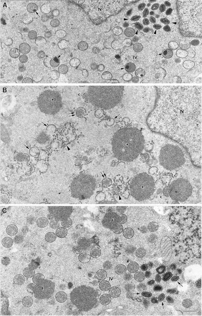
Low-magnification fields of cells infected for 24 h with WR VV or with VVindA14L in the absence or presence of IPTG. (A) IVs, many of them with condensed DNA (arrows), as well as mature virions (arrowheads) accumulate in the cytoplasm of cells infected with WR. (B) The cytoplasm of HeLa cells infected with VVindA14L in the absence of IPTG shows numerous electron-dense masses (asterisks). A few crescent-like structures (c) contact these masses, but most of them do not organize on the surfaces of the masses and accumulate within the cytoplasm (single arrows), as well as membranes that are not organized in crescents (arrowheads). Structures that resemble IVs with interrupted membranes are also seen (double arrows). (C) HeLa cells infected with VVindA14L in the presence of IPTG accumulate characteristic foci of viroplasmic matrix (F) with crescents attached to their surface, IVs, and mature virions (arrows). N, nucleus. Bar, 0.5 μm.