
FIG. 7.
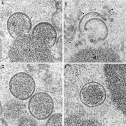
High-magnification fields of the viral crescents and IVs formed in HeLa cells infected for 24 h with VVindA14L in the presence or absence of IPTG. (A) Viral crescents (c) formed on the surfaces of the viroplasmic foci (F) in the presence of IPTG exhibit a thickness and organization indistinguishable from those of the structures generated by the WR VV. (B) The crescent-like structures (arrows) assembled in the absence of IPTG acquire the characteristic curvature of the crescent, but they exhibit a more diffuse appearance. They are usually separated from the surfaces of the dense masses (asterisk), and there are also some membranous elements of irregular shape (arrowheads). (C) IVs from cells infected with the VVindA14L in the presence of IPTG show the same size and apparent organization as the WR VV virions. (D) HeLa cells infected with VVindA14L in the absence of IPTG assemble a few IV-like virions, whose dense internal contents are separated from the membrane of the viral particle (arrows). Bar, 200 nm.