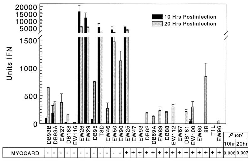
FIG. 4.
Nonmyocarditic viruses induce more IFN-α/β in cardiac myocytes than myocarditic viruses do. Primary cardiac myocyte cultures were infected at an MOI of 5 PFU per cell, and supernatants were removed at 10 and 20 h postinfection. After acidification (to inactivate reovirus) and neutralization of samples and commercial IFN-α/β standards, IFN units were determined by bioassay. P val, P value; MYOCARD, myocarditic potential.

