


FIG. 2.
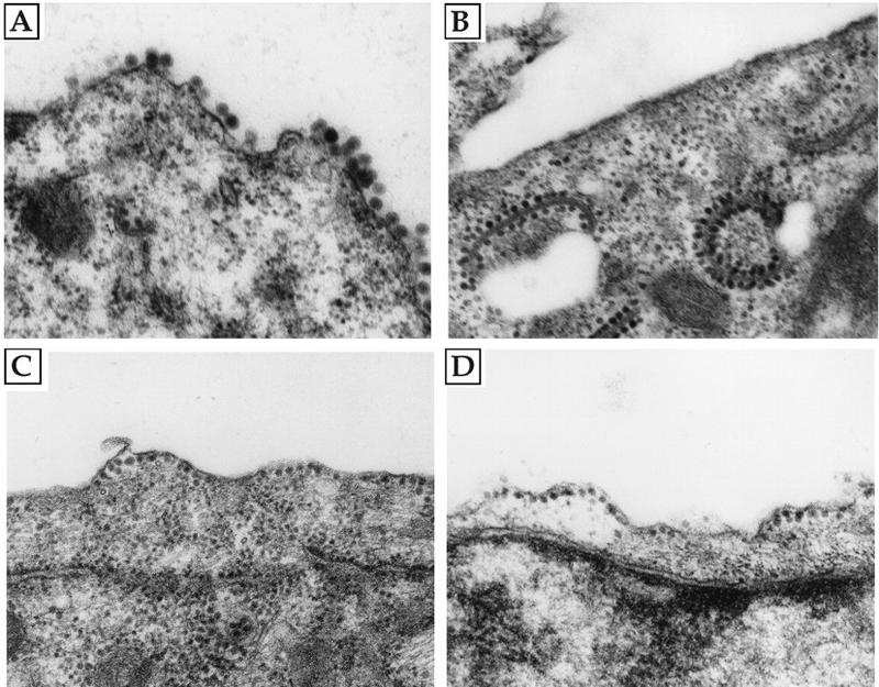
Electron microscopy of infected or transfected cells. BHK cells were transfected with RNA (A, B, and D) or infected with virus (C), and the cells were prepared for electron microscopy after 12 h (A, C, and D) or 24 h (B). (A) Transfection with RNA from pToto54 as a wild-type SIN control; (B) transfection with RNA from the parental chimera pSIN(RRE1); (C) infection with P10 virus from passage series 1; (D) transfection with RNA from the reconstructed adapted chimera pSIN(RRE1)(E2:D248Y/E1:Q411L). Note the abundance of budding virus in panel A and the alignment of nucleocapsids along the plasma membrane in panels C and D, which are not present in panel B.