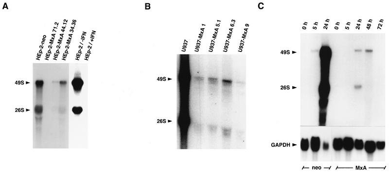
FIG. 5.
Reduced accumulation of SFV genomic 49S and subgenomic 26S RNAs in infected cells expressing MxA protein. Monolayer cultures of different clonal lines of HEp-2-MxA (A) and U937-MxA cells (B) were infected with SFV at an MOI of 3, and total cell RNA was prepared 5 h postinfection. HEp-2-neo and U937 cells served as controls. HEp-2 cells were either left untreated or pretreated with IFN-α2 for 18 h prior to infection (A). (C) To examine RNA accumulation throughout the course of infection, the clonal lines HEp-2-MxA 71.2 and HEp-neo were infected with SFV at an MOI of 1 and total cell RNA was prepared 5, 24, 48, and 72 h postinfection (C). The RNA preparations (20 μg per lane) were subjected to Northern blot analysis with radiolabeled genomic cDNA of SFV. Hybridization to a radiolabeled 0.8-kb fragment of human GAPDH cDNA was used as a control (C, lower section). The blots were exposed to X-ray film.

