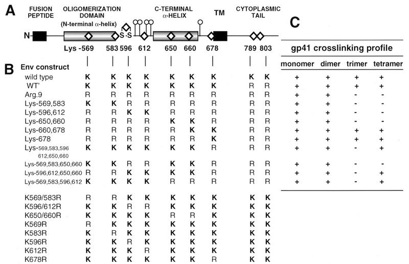
FIG. 1.
(A) Linear diagram of gp41 depicting regions assigned as structural and/or functional domains. The positions of the nine lysine residues targeted with Arg substitutions are indicated by diamonds. Also shown are the N-linked glycosylation sites (○|) and the disulfide loop (S-S). TM, transmembrane sequence. (B) Lys-Arg profiles of gp41 mutants. (C) Summary of mutant gp41 cross-linking profiles (see Fig. 2 to 5) indicating the presence (+) or absence (−) of cross-linked gp41 oligomeric species on immunoblots probed with human α-588-599 antibody.

