
FIG. 2.
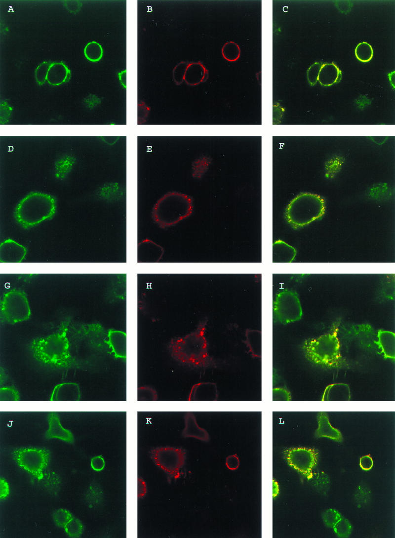
Colocalization of VZV gE and gI during endocytosis. HeLa cells were transfected with the gE gene and the gI gene. The cells were incubated with MAb 6B5 and polyclonal antiserum for gE at 4°C for 30 min. The cells were then returned to 37°C for 0 (A, B, and C), 15 (D, E, and F), 30 (G, H, and I), or 60 (J, K, and L) min. The cells were fixed and permeabilized and then incubated with goat anti-mouse–Texas red conjugate and goat anti-rabbit–FITC conjugate for 1 h. The cells were analyzed by laser scanning confocal microscopy with gE (green stain) (A, D, G, and J) and gI (red stain) (B, E, H, and K); the merged images (yellow stain) are also shown (C, F, I, and L).