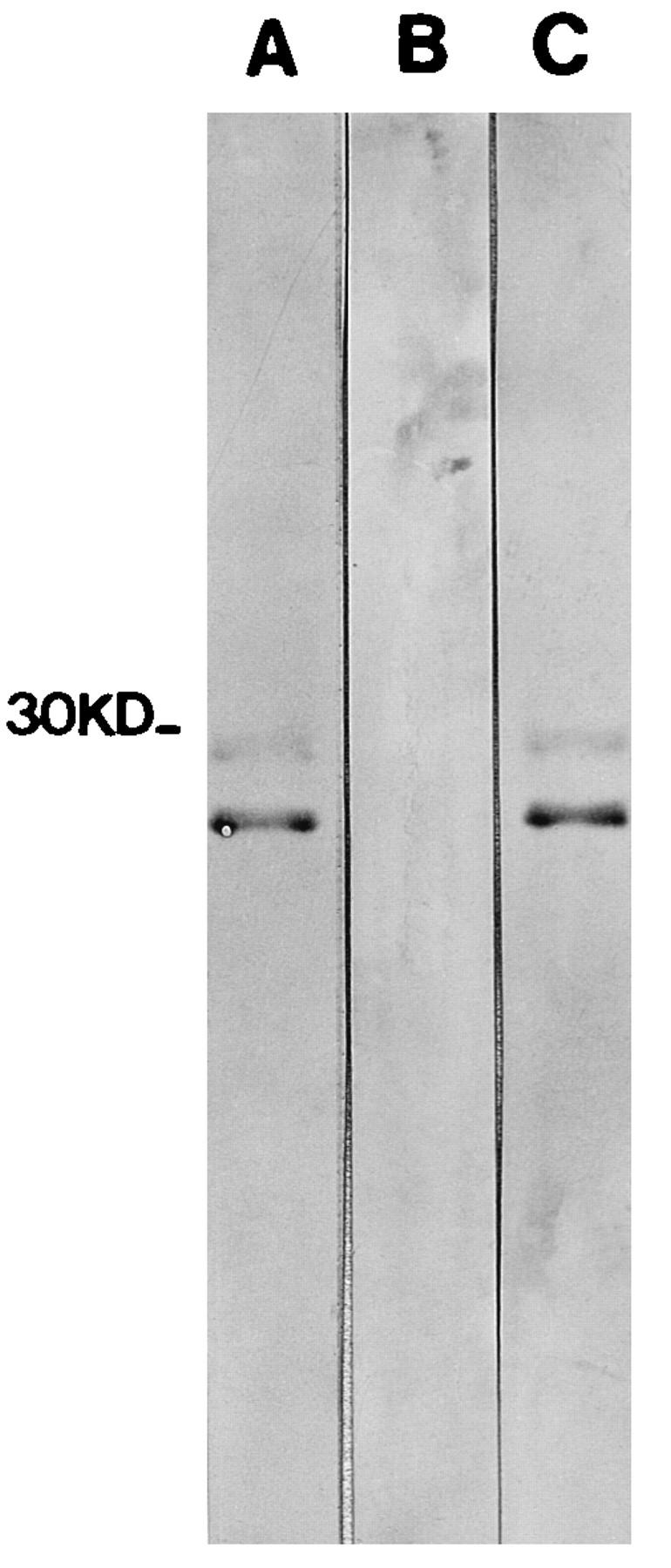
FIG. 4.

Anti-VP1 antibodies detected by Western blotting. FMDV (produced, inactivated, and purified as described by Berinstein et al. [3]) was resuspended in sample buffer (50 mM Tris [pH 7.5], 1 mM phenylmethylsulfonyl fluoride, 4 M urea, 1% sodium dodecyl sulfate [SDS], 2 mM dithiothreitol, and 2% 2-β-mercaptoethanol), boiled 3 min, subjected to SDS–12.5% polyacrylamide gel electrophoresis, and blotted to an Immobilon P (Millipore) membrane. The membrane was blocked overnight with PBST containing 3% skim milk (all subsequent steps were performed with this buffer) and incubated with the corresponding mouse sera (diluted 1/50) for 2 h at 37°C. The membrane was washed and incubated with an alkaline phosphatase-labeled anti-mouse immunoglobulin rabbit antiserum (Dakkopats) for 1 h at 37°C and then washed three more times, and the reaction was developed by the addition of the substrate nitroblue tetrazolium–4-chloro-3-indolylphosphate. Sera from mice immunized with plant extracts were used for the reaction. Lanes A through C correspond to anti-p135-160 antiserum, a pool of sera from mice immunized with pRok-transformed plants, and a pool of sera from mice immunized with pRok.VP1-transformed plants, respectively.
