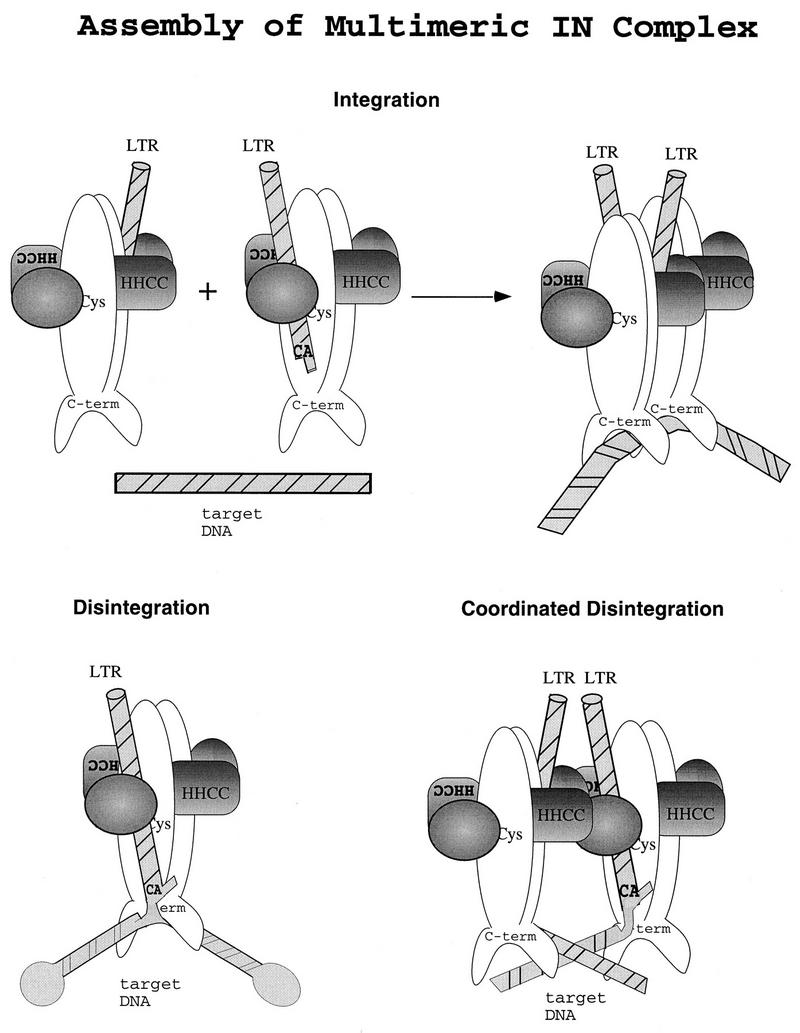
FIG. 6.

Model of multimeric M-MuLV IN functions in vitro and in vivo. An illustration of M-MuLV IN domains and multimeric subunit interactions is shown. A dimeric interface is formed by parallel association of M-MuLV IN monomers, reflecting the structure studies of the catalytic core and C terminus of human immunodeficiency virus type 1 and avian sarcoma virus IN (5, 15, 33). DNA is depicted as striped bars. The N terminus of M-MuLV IN through the HHCC region is shaded. (Top) LTR coordination and multimeric assembly of subunits for integration. (Bottom) Substrate coordination and subunit interactions for unimolecular disintegration (left) and bimolecular coordinated disintegration (right).
