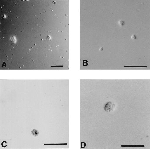
FIG. 2.
Vesicles from amoebae. (A) Low magnification of numerous small vesicles among amoebae (three larger objects) photographed by differential interference contrast microscopy. (B) Higher magnification of three vesicles containing rods, observed by differential interference contrast microscopy. These vesicles were exposed for 3.5 h to MBC 215. (C) Vesicle containing formazan crystals. It was produced by A. polyphaga at 35°C. The light was altered to enhance the visualization of formazan. (D) Unusually large vesicle containing bacteria with formazan crystals. This vesicle was exposed for 4 h at 25°C to MBC 115 and then for 6 h to INT. Bar, 50 (A) and 20 (B through D) μm.

