

Left ventricular pseudoaneurysms are formed when myocardial rupture is contained by pericardial adhesions and thrombus formation. 1 Although rare, false aneurysms of the left ventricle can occur after mitral valve surgery or myocardial infarction. 2 These pseudoaneurysms, contained by visceral pericardium alone, have a tendency to rupture with catastrophic complications. Advances in noninvasive imaging in recent years have resulted in the detection of previously unsuspected pseudoaneurysms. 3 On ultrasound, aneurysms and pseudoaneurysms both exhibit systolic expansion; however, only pseudoaneurysms have rapid flow through the aneurysmal neck during both systole and diastole. 4
A 75-year-old woman with no coronary artery disease underwent mitral valve replacement for severe mitral regurgitation associated with congestive heart failure. A 31-mm pericardial mitral valve was implanted. The procedure was complicated by a ventricular free-wall rupture, 2 × 2 cm in size, which was repaired with a bovine pericardial patch and BioGlue® surgical adhesive (CryoLife International®, Inc.; Kennesaw, Ga). An echocardiogram performed 2½ months after surgery did not reveal any abnormal shunting. However, 2 weeks later, the patient's heart failure worsened, and cardiac echocardiography revealed dehiscence of the patch (Figs. 1–3). The patient returned to surgery for patch repair. The pericardial space was found to be obliterated by adhesions, and the pseudoaneurysm was filled with a large amount of thrombus. The patch and the BioGlue had completely separated from the myocardium, leaving a 4- × 6-cm defect. The previous patch and the BioGlue were removed. New patches were placed on the epicardial and endocardial sides of the myocardial defect (double-patch repair). The procedure was successful, and the patient was discharged from the hospital doing well.
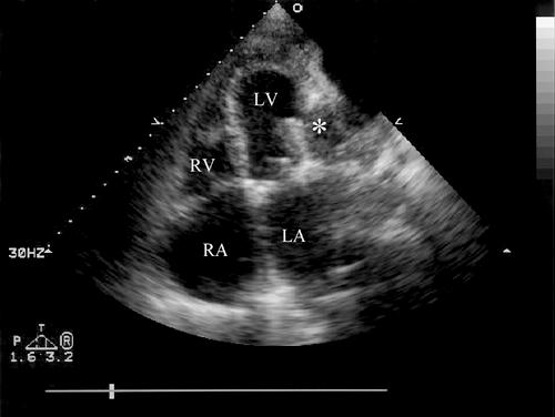
Fig. 1 Apical 4-chamber view of the echocardiogram shows the rupture with lack of myocardium in the lateral wall of the left ventricle, and the cavity of the pseudoaneurysm (*).
LA = left atrium; LV = left ventricle; RA = right atrium; RV = right ventricle

Fig. 2 A color Doppler echocardiogram, off-axis 4-chamber view, shows a broad jet of color in the neck of the pseudoaneurysm, indicating blood flow into the cavity.

Fig. 3 A pulse-wave Doppler from the apical 4-chamber view of the echocardiogram reveals flow during both systole and diastole, which is pathognomonic of a pseudoaneurysm.
Footnotes
Address for reprints: Ming Hui Chen, MD, MMSc, Associate Director, Noninvasive Cardiac Laboratory, Brigham and Women's Hospital and Harvard Medical School, 75 Francis Street, Boston, MA 02115
References
- 1.Frances C, Romero A, Grady D. Left ventricular pseudoaneurysm. J Am Coll Cardiol 1998;32:557–61. [DOI] [PubMed]
- 2.Chan R, Lubicz S, Oliver L, Calafiore P. Left ventricular false aneurysm complicating mitral valve repair. Ann Thorac Surg 1993;56:175–6. [DOI] [PubMed]
- 3.Natarajan MK, Salerno TA, Burke B, Chin B, Armstrong PW. Chronic false aneurysms of the left ventricle: management revisited. Can J Cardiol 1994;10:927–31. [PubMed]
- 4.Wiegers SE. Left ventricular pseudoaneurysm. In: Wiegers SE, Plappert T, St John Sutton M, editors. Echocardiography in practice: a case-oriented approach. Malden (Mass): Blackwell Science; 2001. p. 269–70.