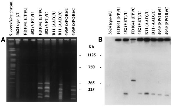
FIG. 2.
PFGE evidence supporting the localization of cpe to episomes in isolates from some disease sources. PFGE and Southern hybridization analysis of undigested and I-CeuI-digested genomic DNA from select C. perfringens disease isolates. (A) pulsed-field gel stained with ethidium bromide. (B) Southern blot of the gel shown in panel A. The blot was probed with a 639-bp DIG-labeled cpe-specific fragment. FP, food poisoning; VET, veterinary; U, undigested, intact genomic DNA; C, I-CeuI-digested DNA. The gel was calibrated with Saccharomyces cerevisiae chromosomal DNA. Molecular sizes of the DNA markers are given in the center. See Table 1 for full strain designations.

