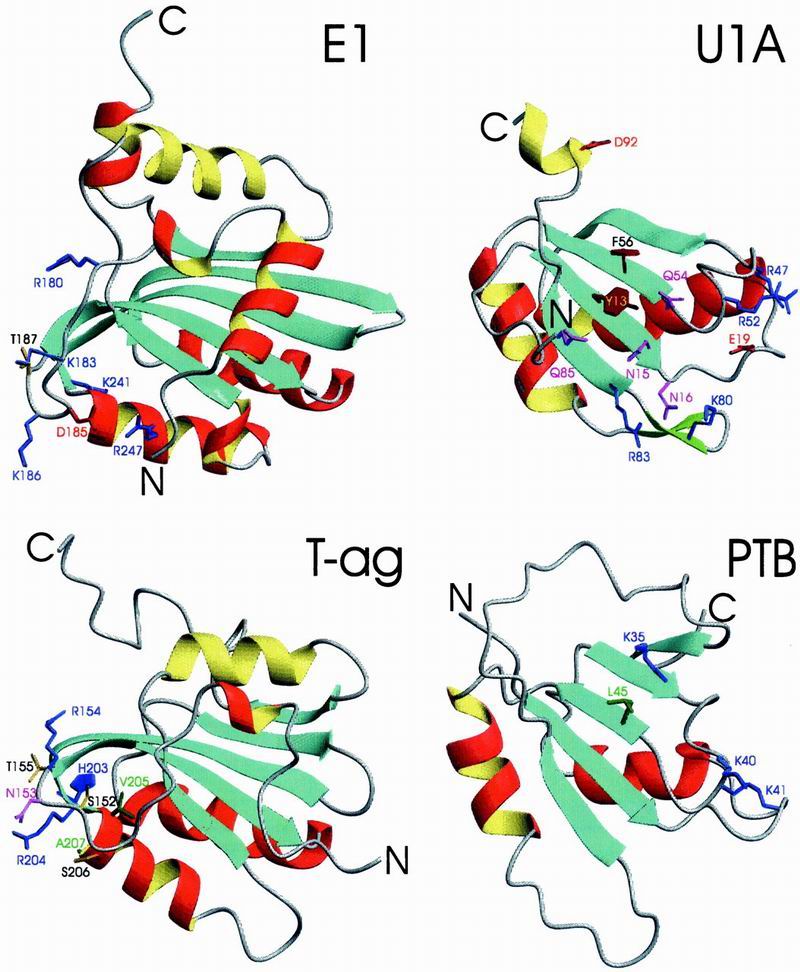
Fig 4.

Ribbon representation of the DBDs of papillomavirus protein E1 and SV40 T-ag and RBDs of U1A and PTB. Helices conserved between Rep and D/RBDs are colored in red (α1) and yellow (α2), all other helices in red/yellow (outside/inside), strands in blue and loops and chain termini in gray. Side chains of amino acids implicated in nucleic acid recognition are displayed and colored identically to the color code given for Fig. 2C. PDB accession codes are E1, ; U1A, ; T-ag, ; and PTB, .
