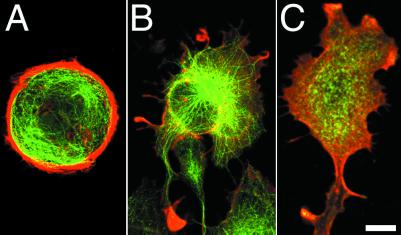
Fig 6.
Localization of actin filaments, microtubules, and myosin-II in IAR-2 epitheliocytes treated with Y-27632. Cells were double-stained for actin (red) and tubulin (green) (A and B). Control cells not treated with inhibitor possessed thick marginal bundles of actin filaments that isolated microtubules from the edge (A). Upon treatment with Y-27632, marginal bundles disassembled, and microtubules radiated out to the cell edges and into processes (B). Double-labeling cells incubated with Y-27632 for actin (red) and nonmuscle myosin-II (green) established the absence of myosin-II or actin filament containing stress fibers (C). Actin filaments were present at the edges of cells; however, myosin-II tended to be diffusely localized throughout the cell (C). (Bar = 10 μm.)

