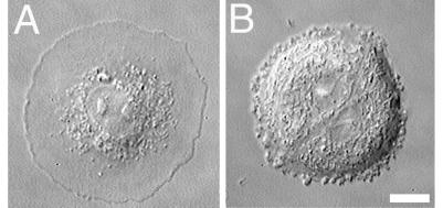
Fig 7.
Video-differential interference contrast micrographs of epithelial IAR-2 cells treated with nocodazole and Y-27632 or with nocodazole. Cell treated with 3 μM nocodazole and 50 μM Y-27632 for 5 h became highly flattened and discoid shaped (A). Note the absence of organelles within the flattened lamella. Group of two rounded cells treated with nocodazole for 5 h formed small swellings at the edges (B). (Bar = 10 μm.)

