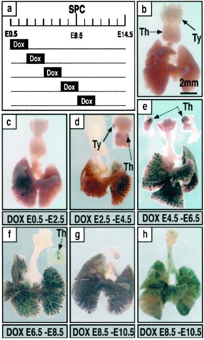
Fig 5.
Temporal specificity of recombination. Dams were treated with doxycycline for 48 h and labeling assessed at E14.5 by whole-mount AP staining. A diagram of the treatment protocols used during embryonic development is shown (a). E0.5 is 12 h after fertilization as determined by detection of a vaginal plug. Whole-mount AP staining of lungs from triple-transgenic mice was assessed on E14.5 to determine the extent of recombination (b–h). Single-transgenic control littermate (b). Partial lobar labeling in the peripheral lung after doxycycline from E0.5 to E2.5 (c). Extensive labeling of intrapulmonary tissue with variable staining in the thymus and thyroid seen among littermates (d). Labeling of most intrapulmonary epithelial cells. Staining was observed in the thymus of all mice but not in the thyroid (e). Labeling in the peripheral lung was found in all mice; recombination in the thymus was found frequently but not in all mice (f). Labeling of extrapulmonary airways was never observed from E0.5to E8.5 (c–f). From E8.5 to E10.5, labeling of intrapulmonary epithelial cells was widespread or focal with variation seen among littermates; subsets of cells in extrapulmonary airways were labeled, the numbers of labeled cells being more abundant in the periphery, and labeling was not seen in thymus or thyroid (g and h). Extensive, but not complete labeling of intrapulmonary epithelial cells was observed in association with labeling of some cells in the extrapulmonary airways (h). (Bar = 2 mm.) Th, thymus; Ty, thyroid.

