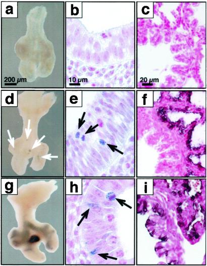
Fig 6.
Progenitors of the peripheral lung are highly restricted at E11.5. Lungs were obtained at E11.5 (a, b, d, e, g, and h), at E18.5 (c, f, and i), from control mice (a–c) or triple-transgenic mice SP-C-rtTA/(tetO)Cre/Zap (d, f, g, and i) and SP-C-rtTA/(tetO)Cre/ZEG (e and h) after doxycycline treatment from E6.5 to E8.5 (d–f) or E8.5 to E10.5 (g–i). No AP or GFP staining was seen in controls. At E11.5, AP-labeled cells seen after treatment with doxycycline from E6.5 to E8.5 were highly restricted to small clusters along the bronchial tubes and to the tips of the lobar bronchi (d). At higher magnification, small clusters of GFP-immunostaining cells were noted at these locations (e). This same period of doxycycline exposure resulted in nearly complete labeling of peripheral lung with exclusion of large conducting and extrapulmonary airways at E18.5 (f). After doxycycline exposure, from E8.5 to E10.5, more extensive AP (g) or GFP (h) was seen in proximal and peripheral lung tubules at E11.5. Consistent with the more widespread labeling of extrapulmonary conducting airways seen at E18.5, patchy labeling of the peripheral lung parenchyma was also noted at E18.5 (i). [Bars = 200 μm, (a, d, g), 10 μm, (b, e, h), and 20 μm (c, f, i).]

