
Figure 1.
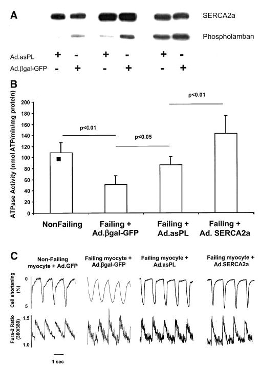
A, Phospholamban and SERCA2a expression from 3 different isolated cardiomyocyte preparations 48 hours after infection with Ad.asPL or Ad.βgal-GFP. B, Measurements of ATPase activity performed at [Ca2+] of 10 μmol/L in membrane preparations from nonfailing cardiomyocytes (n = 10) and failing cardiomyocytes infected for 48 hours with Ad.asPL (n = 6), Ad.SERCA2a (n = 8), or Ad.βgal-GFP (n = 8). C, Recordings from cardiomyocytes isolated from a donor nonfailing heart and from a failing heart infected with an adenovirus expressing either Ad.βgal-GFP, Ad.asPL, or Ad.SERCA2a, stimulated at 1 Hz at 37°C. The failing cells had a characteristic decrease in contraction and prolonged relaxation, along with a prolonged Ca2+ transient. Ablation of phospholamban in the failing cardiomyocytes normalized these parameters.