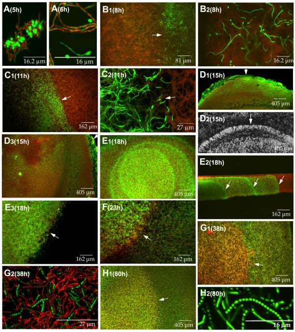
Figure 1.
Confocal laser-scanning fluorescence microscopy analysis of the development-linked cell death processes of Streptomyces antibioticus ATCC11891 in confluent surface cultures. Developmental phases (A-H) and culture times (hours) are indicated. Picture D2 was obtained under the phase contrast microscope. The other images correspond to culture sections stained with SYTO 9 and propidium iodide. E2 is a cross section view; the other images correspond to longitudinal sections (see methods). Arrows in E2 indicate the eccentric circles of live mycelium developing from the bottom upwards, forming distinct layers with well-defined boundaries. Arrows in the rest of the images indicate circle edges. For details see text.

