Figure 3.

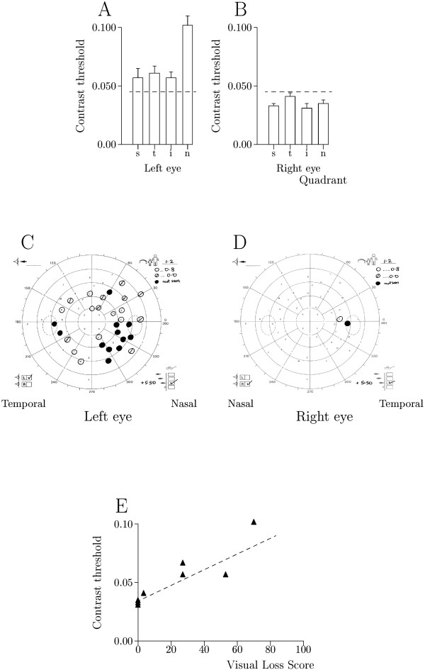
Patient 9 (Table 1) A & B Histograms of contrast threshold (mean S.E.M.) for superior (s), temporal (t), inferior (i) and nasal (n) truncated quadrants in the left and right eyes showing upper 95% prediction limit of 0.045 contrast units as broken horizontal line. Contrast thresholds were abnormal in the left eye. C & D Friedmann visual fields showing: unmarked letters-normal detection (1.2 logarithmic units attenuation), reduced sensitivity (○ 0.8 logarithmic units attenuation and ø zero attenuation) and ● zero detection. The main visual field loss was in the inferior hemifield of the left eye. E Contrast threshold against visual field loss score taken from Friedmann charts for each truncated quadrant, showing best fitting relationship as broken line. Relationship was significant: R2 = 85%, p = 0.001.
