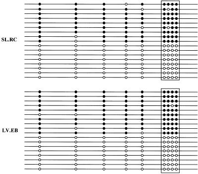
Fig 2.
Normal half-methylation of a CTCF binding site in the H19 ICR, analyzed by bisulfite DNA sequencing. PCR products were cloned and 20 randomly selected clones were sequenced for each EG line. Methylated CpG sites are depicted by filled circles, and the unmethylated CpG sites as open circles. The boxed area represents the CTCF core binding site 1 (ref. 10). (Upper) Line SL.RC. (Lower) Line LV.EB.

